Quick Order Cart

Amyotrophic Lateral Sclerosis (ALS) Research

Home / Applications / Area of Research Interest / ALS Research and Cell Models
Neuroscience Research

ALS Research and Cell Models

Cell models for studying motor neuron degeneration, glial toxicity, neuroinflammation, neuromuscular junction impairment, and blood–CNS barrier dysfunction.

ALS research and cell models
Motor Neuron Degeneration
Glial Toxicity
NMJ Dysfunction
Barrier Models
01

Motor Neuron Degeneration

Study axonal dysfunction, excitotoxicity, oxidative stress, and ALS-associated molecular pathways.

02

Glial Contribution

Model astrocyte- and microglia-mediated effects on motor neuron survival and inflammatory signaling.

03

NMJ Impairment

Explore motor neuron–muscle communication, denervation, myotube response, and muscle atrophy.

04

Blood–CNS Barrier

Investigate endothelial cells, pericytes, tight junctions, and blood–spinal cord barrier dysfunction.

Introduction

Understanding ALS Through Cell-Based Research

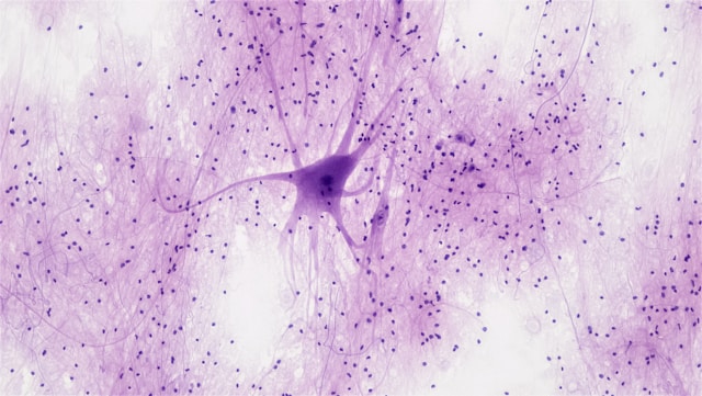
Amyotrophic lateral sclerosis (ALS) is a fatal neurodegenerative disorder of the human motor system. It is characterized by the selective and progressive degeneration of upper motor neurons in the motor cortex and lower motor neurons in the brainstem and anterior horn of the spinal cord. Loss of upper motor neurons commonly leads to spasticity, hyperreflexia, and impaired voluntary motor control, whereas degeneration of lower motor neurons is associated with muscle weakness, fasciculations, muscle wasting, and eventual paralysis. Clinically, ALS often begins with focal muscle weakness or twitching in an arm or leg, difficulty swallowing, or slurred speech. As the disease progresses, it increasingly affects movement, speech, swallowing, nutrition, and respiration.

The etiology of ALS remains incompletely understood, and no curative treatment is currently available. In ALS, progressive motor neuron degeneration weakens or disrupts the transmission of nerve signals to skeletal muscles. As a result, affected muscles lose neural stimulation, become progressively weaker, undergo atrophy, and eventually fail to function properly. Approximately 10% of ALS cases are familial, many of which are associated with pathogenic mutations in genes involved in neuronal function, RNA processing, protein homeostasis, oxidative stress, or axonal maintenance. However, most ALS cases are sporadic, and the mechanisms driving disease onset and progression remain poorly defined. Despite extensive research, current clinical management remains limited. Existing therapies primarily aim to slow disease progression, modestly extend survival, preserve function, manage symptoms, and improve quality of life, but they generally do not reverse neuronal loss once motor neurons have degenerated.

ALS research increasingly relies on cell-based models to study both motor neuron-intrinsic degeneration and non-cell-autonomous mechanisms involving glial cells, skeletal muscle, peripheral nerve support cells, and vascular barrier components. These models help researchers investigate disease mechanisms, evaluate pathway-specific responses, and develop experimental systems for preclinical discovery.

Need Help Choosing?

Looking for Cell Models for ALS Research?

Tell us your research focus, target species, preferred culture format, and experimental design. Our team can help recommend suitable cell models and culture systems.

Research Strategy

Why Cell Models Matter in ALS Research

ALS is not only a motor neuron disease at the cellular level. Although motor neurons are the primary affected cells, disease progression involves multiple interacting cell types across neuronal, glial, muscular, peripheral nerve, and vascular systems.

N

Neuronal Mechanisms

Study motor neuron degeneration, axonal dysfunction, excitotoxicity, mitochondrial stress, and ALS-associated gene pathways.

G

Glial and Inflammatory Mechanisms

Use astrocytes, microglia, and oligodendrocyte lineage cells to study non-cell-autonomous toxicity and neuroinflammation.

V

Muscle and Vascular Systems

Explore neuromuscular junction impairment, muscle atrophy, endothelial barrier dysfunction, and pericyte-related vascular changes.

Cell Type Selection

ALS-Related Cell Types and Research Applications

The following cell types are commonly associated with ALS-related research areas, including motor neuron degeneration, glial toxicity, neuroinflammation, neuromuscular junction impairment, and barrier dysfunction.

Cell Type ALS Research Focus References
Motor Neurons / Spinal Motor Neurons Core ALS disease modeling; motor neuron degeneration; axonal dysfunction; excitotoxicity; SOD1, C9orf72, TARDBP, and FUS-related mechanisms. Brown & Al-Chalabi, 2017, New England Journal of Medicine; Taylor et al., 2016, Nature.
Astrocytes Non-cell-autonomous toxicity; astrocyte-mediated motor neuron injury; glutamate metabolism; oxidative stress; inflammatory signaling. Di Giorgio et al., 2008, Cell Stem Cell; Stoklund Dittlau & Van Den Bosch, 2023, Frontiers in Molecular Medicine.
Microglia Neuroinflammation; microglial activation; inflammatory cytokine release; microglia–motor neuron interaction; disease progression. Clarke & Patani, 2020; Bond et al., 2025.
Oligodendrocytes / OPCs Myelin and axonal metabolic support; oligodendrocyte-mediated motor neuron injury; OPC differentiation; MCT1/lactate support. Ferraiuolo et al., 2016, PNAS; Jamet et al., 2024.
Skeletal Muscle Cells / Myoblasts / Myotubes Neuromuscular junction impairment; muscle atrophy; motor neuron–muscle co-culture; muscle-side contribution to ALS pathology. Campanari et al., 2016; Shefner et al., 2023, Brain.
Schwann Cells Peripheral nerve support; terminal Schwann cells; NMJ stability; peripheral contribution to ALS progression. Moss et al., 2025; Lewis et al., 2025.
Brain / Spinal Cord Microvascular Endothelial Cells BBB/BSCB dysfunction; blood-spinal cord barrier breakdown; tight junction disruption; vascular contribution to ALS. Zhong et al., 2008; Steinruecke et al., 2023.
Pericytes Blood-spinal cord barrier integrity; pericyte loss; barrier leakage; vascular niche changes in ALS. Winkler et al., 2013; Steinruecke et al., 2023.
Featured Research Areas

Model ALS from Multiple Biological Angles

Researchers can select cell models based on the mechanism being studied, from motor neuron degeneration to glial contribution, neuromuscular dysfunction, and neurovascular impairment.

Motor Neuron Degeneration

Motor neuron models are used to study axonal degeneration, excitotoxicity, mitochondrial dysfunction, protein aggregation, oxidative stress, and ALS-associated genetic pathways.

Motor Neurons Spinal Neurons Neural Progenitor Models

Glial Contribution to ALS

Astrocytes, microglia, and oligodendrocyte lineage cells may influence motor neuron survival through inflammatory signaling, glutamate regulation, metabolic support, and oxidative stress.

Astrocytes Microglia Oligodendrocytes OPCs

Neuromuscular Junction and Muscle Involvement

Skeletal muscle cells, myoblasts, myotubes, and Schwann cells can be used to study NMJ disruption, denervation, muscle atrophy, and peripheral contributions to disease progression.

Skeletal Muscle Cells Myoblasts Myotubes Schwann Cells

Blood–CNS Barrier Dysfunction

Endothelial cells and pericytes can be used to investigate barrier integrity, tight junction disruption, vascular leakage, and neurovascular interactions in ALS-related research.

Brain MVECs Spinal Cord MVECs Pericytes Barrier Co-culture
Recommended Categories

Explore Cell Models and Supporting Products

Select products by research focus, cell type, tissue origin, culture system, or supporting reagent needs.

Neural and Glial Cells

  • Motor neurons and spinal neurons
  • Astrocytes and microglia
  • Oligodendrocytes and OPCs
  • Neural progenitor cells
Explore Neuroscience Models

Neuromuscular and Peripheral Models

  • Skeletal muscle cells
  • Myoblasts and myotubes
  • Schwann cells
  • Motor neuron–muscle co-culture support
Browse Primary Cells

Vascular and Barrier Models

  • Brain microvascular endothelial cells
  • Spinal cord microvascular endothelial cells
  • Pericytes
  • BBB/BSCB-related culture systems
View Barrier-Related Cells

Immortalized Cell Lines

  • Stable and scalable in vitro systems
  • Neuroinflammation-related cell lines
  • Repeatable assay development
Browse Immortalized Cells

Genome-Edited Cells

  • Knockout cell lines
  • Overexpression cell lines
  • Reporter cell lines
  • Pathway-specific studies
Browse Genome-Edited Cells

Media and Reagents

  • Neuronal culture media
  • Astrocyte and microglia media
  • Endothelial cell media
  • Coating reagents and supplements
Explore Media and Reagents
Selection Guide

How to Choose a Cell Model for ALS-Related Studies

Different ALS-related mechanisms require different cell model strategies. Start with the biological question, then select the cell type, species, culture format, and supporting reagents.

Define the disease mechanism

Motor neuron degeneration, glial toxicity, neuroinflammation, NMJ dysfunction, or barrier impairment.

Select the relevant cell system

Choose neuronal, glial, muscular, peripheral nerve, or vascular cell models based on your study goal.

Consider co-culture and assay design

Use co-culture systems when studying cell–cell interactions such as neuron–astrocyte or motor neuron–muscle communication.

Match media and culture support

Confirm culture media, supplements, coating reagents, and handling conditions before starting experiments.

Why Ascent Research

Cell Culture Support for ALS-Related Research

Quality-Certified

Cell models and related products supported by quality control and documentation where applicable.

Fast-Delivered

Responsive sourcing and delivery support to help researchers move projects forward efficiently.

$

Budget-Friendly

Flexible product options for academic labs, biotech teams, and early-stage discovery programs.

?

Expert-Backed

Technical support for product selection, culture conditions, and cell model planning.

Build Your ALS Research Model with Ascent Research

Whether your study focuses on motor neuron degeneration, glial toxicity, neuroinflammation, neuromuscular junction dysfunction, or vascular barrier impairment, Ascent Research can help you identify suitable cells, media, and culture support products.

FAQ

Frequently Asked Questions

What cell type is most directly relevant to ALS research?

Motor neurons are the most directly relevant cell type because ALS is primarily characterized by progressive degeneration of upper and lower motor neurons. However, ALS progression also involves glial, muscular, peripheral nerve, and vascular components.

Are astrocytes and microglia useful for ALS studies?

Yes. Astrocytes and microglia are widely used to study non-cell-autonomous mechanisms in ALS, including neuroinflammation, glutamate metabolism, oxidative stress, and inflammatory cytokine signaling.

Can skeletal muscle cells be used in ALS-related research?

Yes. Skeletal muscle cells, myoblasts, and myotubes can be used to study muscle atrophy, denervation, neuromuscular junction dysfunction, and motor unit degeneration.

Are endothelial cells and pericytes relevant to ALS?

Yes. Brain and spinal cord microvascular endothelial cells, together with pericytes, can be used to study blood–brain barrier and blood–spinal cord barrier dysfunction in ALS-related research.

Can Ascent Research help select suitable cell models?

Yes. Ascent Research can help researchers select cell models based on research focus, species, tissue origin, culture format, and experimental design.

References

Selected References

  1. Feldman, E. L., Goutman, S. A., Petri, S., Mazzini, L., Savelieff, M. G., Shaw, P. J., & Sobue, G. 2022. Amyotrophic Lateral Sclerosis. The Lancet 400(10360):1363–1380. doi:10.1016/S0140-6736(22)01272-7.
  2. Kiernan, M. C., Vucic, S., Cheah, B. C., Turner, M. R., Eisen, A., Hardiman, O., Burrell, J. R., & Zoing, M. C. 2011. Amyotrophic Lateral Sclerosis. The Lancet 377(9769):942–955. doi:10.1016/S0140-6736(10)61156-7.
  3. Stoklund Dittlau, K., & Van Den Bosch, L. 2023. Why Should We Care about Astrocytes in a Motor Neuron Disease? Frontiers in Molecular Medicine 3:1047540. doi:10.3389/fmmed.2023.1047540.
  4. Brown, R. H., & Al-Chalabi, A. 2017. Amyotrophic Lateral Sclerosis. New England Journal of Medicine.
  5. Taylor, J. P., Brown, R. H., & Cleveland, D. W. 2016. Decoding ALS: From Genes to Mechanism. Nature.
  6. Di Giorgio, F. P., et al. 2008. Human Embryonic Stem Cell-Derived Motor Neurons Are Sensitive to the Toxic Effect of Glial Cells Carrying an ALS-Causing Mutation. Cell Stem Cell.
  7. Ferraiuolo, L., et al. 2016. Oligodendrocytes Contribute to Motor Neuron Death in ALS via SOD1-Dependent Mechanism. Proceedings of the National Academy of Sciences.
  8. Campanari, M.-L., et al. 2016. Neuromuscular Junction Impairment in Amyotrophic Lateral Sclerosis. Frontiers in Molecular Neuroscience.
  9. Shefner, J. M., et al. 2023. Skeletal Muscle in Amyotrophic Lateral Sclerosis. Brain.
  10. Winkler, E. A., et al. 2013. Blood-Spinal Cord Barrier Breakdown and Pericyte Reductions in Amyotrophic Lateral Sclerosis. Acta Neuropathologica.
  11. Zhong, Z., et al. 2008. ALS-Causing SOD1 Mutants Generate Vascular Changes Prior to Motor Neuron Degeneration. Nature Neuroscience.
  12. Steinruecke, M., et al. 2023. Blood-CNS Barrier Dysfunction in Amyotrophic Lateral Sclerosis. Journal of Cerebral Blood Flow & Metabolism.
Research Use Only: All products and services described on this page are intended for research use only. They are not intended for diagnostic, therapeutic, clinical, or human use.
Reset Password

    Reach Us Questions? Click Me Here!

    Fill out the form below and a member of our team will contact you shortly!

    *Required field


    Referee Details Discounts will be issued to both accounts after manual review!

      🎁 Refer & Earn · Rewards for You & Your Friends

      ✨ Your Referral Link