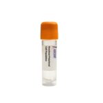
The LYPD3 Knockout Raji Polyclonal Cells are a CRISPR/Cas9-edited polyclonal knockout cell population derived from the human Raji B lymphocyte line. This product provides a heterogeneous pool of cells carrying disruptions in the LYPD3 gene, generated via CRISPR/Cas9-mediated gene editing to create a loss-of-function model for studying LYPD3-dependent cellular processes.
Raji cells are a suspension B lymphocyte line derived from a Burkitt lymphoma patient, known for their Epstein-Barr virus (EBV)-positive status and robust antibody production and antigen presentation capabilities. Their rapid growth and ease of genetic manipulation make them an ideal host for generating CRISPR/Cas9-edited knockout populations to study signaling pathways in a hematopoietic context.
LYPD3 (C4.4A) is a GPI-anchored cell surface glycoprotein that promotes cell adhesion, migration, and invasion. It binds laminin-5 and cooperates with integrin ??3??1 to activate FAK and Src, leading to cytoskeletal reorganization. Upstream, LYPD3 is regulated by TGF-??, Wnt/??-catenin, and AP-1. Downstream effectors include PI3K/AKT and MMPs. This signaling axis positions LYPD3 as a critical mediator of focal adhesion dynamics and integrin-mediated signal transduction, contributing to enhanced cell motility and metastatic dissemination.
In the Raji B lymphocyte background, LYPD3 knockout cells enable dissection of LYPD3 functions outside epithelial cancer models. Although Raji cells grow in suspension, they express integrins and can be stimulated to adhere and migrate upon ECM engagement. This polyclonal knockout population facilitates study of LYPD3-dependent signal transduction, such as FAK and Src activation, independent of epithelial adhesion complexes. It offers a model for investigating how LYPD3 influences migration and invasion signaling in hematopoietic cells, potentially informing lymphoma dissemination or immune cell trafficking.
Applications include cancer metastasis modeling, cell adhesion and migration studies, and anti-metastatic drug screening. The knockout cells enable Western blotting for LYPD3, FAK, and phospho-FAK, as well as migration and invasion assays. Immunofluorescence for focal adhesion markers and RT-qPCR for EMT markers are also suitable. These cells provide a platform to study the LYPD3-integrin ??3??1-laminin-5 interaction and to screen small molecules targeting LYPD3-driven pathways. For technical inquiries, please contact Ascent Research.