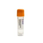
The MFSD8 Knockout Raji Polyclonal Cells are a CRISPR/Cas9-edited polyclonal knockout cell population targeting MFSD8 in Raji B lymphocytes. This polyclonal model provides a heterogeneous population with MFSD8 gene disruption, enabling the study of lysosomal dysfunction in a human Burkitt??s lymphoma background and preserving genetic diversity for autophagy and lysosomal storage disease research.
Raji cells are an EBV-positive human B lymphocyte line derived from a Burkitt??s lymphoma patient, widely used as a model for B cell malignancies and immune studies. Their mature B cell phenotype, including antigen-presenting capacity, makes them suitable for investigating lysosomal dynamics in the context of immune cell biology and oncogenic signaling.
MFSD8 encodes a lysosomal transmembrane protein that functions as a transporter central to lysosomal homeostasis and autophagy. Its transcription is activated by TFEB and TFE3, downstream of mTORC1 inhibition during starvation or lysosomal stress. MFSD8 interacts with other late-infantile neuronal ceroid lipofuscinosis proteins, including CLN3, CLN5, CLN6, and CLN8, as well as v-ATPase subunits, to regulate lysosomal acidification. Downstream, MFSD8 promotes autophagic flux and cathepsin-mediated degradation; its disruption causes accumulation of autophagic substrates LC3-II and p62/SQSTM1, defective mTORC1 reactivation, and lysosomal dysfunction characteristic of CLN7 disease.
In the Raji B lymphocyte model, MFSD8 knockout enables dissection of lysosomal pathways in an immune context. Lysosomes in B cells are essential for antigen processing, MHC class II presentation, and signal transduction; thus, impaired autophagy and lysosomal exocytosis due to MFSD8 loss may perturb immune function and mimic neurodegenerative lysosomal storage pathology. This model facilitates studies on how lysosomal stress influences B cell proliferation, survival, and oncogenic signaling, bridging cancer biology and Batten disease research.
Researchers can utilize these polyclonal knockout cells for mechanistic autophagy studies, drug screening to identify lysosomal modulators or autophagy inducers, and functional mapping of the CLN7 protein interaction network. Representative experimental techniques include quantitative Western blotting for MFSD8, LC3B, and p62; RT-qPCR analysis of lysosomal genes such as LAMP1 and LAMP2; immunofluorescence staining for lysosomal membrane markers; LysoSensor-based lysosomal pH assessment; transmission electron microscopy to visualize storage deposits; and flow cytometry to measure cell viability and apoptosis. For additional details, please contact Ascent Research.