Quick Order Cart

Cat. No. ARG1510

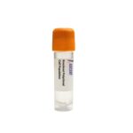
MICAL2 Knockout Raji Polyclonal Cells

  • Product Type:

    Polyclonal Cell Population

  • Species:

    Homo sapiens (Human)

  • Tissue Source:

    Bone

  • Disease:

    Burkitt lymphoma

This product provides a CRISPR/Cas9-edited polyclonal knockout cell population targeting MICAL2 in the Raji B lymphoblastoid cell line, a well-characterized model for B cell biology and lymphoma research. MICAL2 oxidizes actin methionine-44 to trigger F-actin depolymerization, functioning downstream of semaphorin-plexin signaling to regulate migration, adhesion, and cytoskeletal organization. By eliminating MICAL2 expression, these polyclonal knockout cells enable functional dissection of actin remodeling pathways in lymphocyte migration and tumor cell invasion. Typical assays include transwell migration, Matrigel invasion, immunofluorescence with phalloidin, and drug screening for anti-metastatic agents. This model is valuable for semaphorin pathway analysis and cytoskeletal drug testing.

Inquire Now

In stock

Ships next business day


Ask a Question

Shipping Info:

Cryopreserved in vials and shipped on dry ice


Disclaimer:

For Research Use Only

  • Characteristics

    Host Cell

    Raji

    Cell Type

    B cell line

    Sex of Donor

    Male

    Age

    11 years

    Derived From Site

    In situ; Maxilla

    Gene Name

    MICAL2

    Gene Identifier

    NCBI Gene ID 9645

    Morphology

    Lymphoblast-like

    Growth Mode

    Suspension

    Storage

    Liquid nitrogen (LN2)

  • Culture Conditions

    Growth medium

    RPMI 1640

    Supplement(s)

    10% Fetal Bovine Serum, 1% Penicillin-Streptomycin Solution

    Temperature

    37°C

    Atmosphere

    5% COâ‚‚

  • Quality Control

    Sterility testing

    The bacterial, yeast, and fungi are not detected in these cells by daily monitor.

    Mycoplasma testing

    Negative for mycoplasma through PCR analysis

  • Disclaimer

    Intended Use

    This product is intended for laboratory in vitro use only. It is not intended for diagnostic, therapeutic, or clinical applications.

    Disclaimer

    Ascent Research endeavors to provide accurate and up-to-date product information. However, no warranties or representations are made regarding its completeness or reliability. References to scientific literature and patents are for informational purposes only, and the customer assumes sole responsibility for verifying their accuracy.

    By accepting this product, the customer acknowledges and agrees to assume all risks associated with its receipt, handling, storage, disposal, and use, including compliance with all applicable safety and environmental regulations and precautions. Relevant laws, regulations, and ethical guidelines must be followed in conducting any research, modifications, or derivatives derived from this product.

    This product is provided "AS IS", and except as expressly stated herein, Ascent Research disclaims all other warranties, express or implied. Under no circumstances shall Ascent Research, its affiliates, or representatives be liable for indirect, incidental, consequential, or punitive damages arising from the use of this material. While Ascent Research employs rigorous quality control measures, we shall not be held responsible for damages resulting from misidentification or misinterpretation of the provided materials.

Description

This MICAL2 Knockout Raji Polyclonal Cells product comprises a CRISPR/Cas9-edited polyclonal population of Raji B lymphoblastoid cells carrying targeted disruption of the MICAL2 gene. The polyclonal format provides a heterogeneous loss-of-function model that avoids clonal selection bias, enabling robust studies of MICAL2-dependent actin remodeling and semaphorin signaling in a lymphoma-derived background. This knockout pool is designed for functional genomics, drug screening, and mechanistic studies in B-cell biology.

Raji is an EBV-positive B lymphocyte cell line originally established from a Burkitt lymphoma patient. It is extensively used to dissect B cell receptor signaling, antigen processing and presentation, and apoptosis pathways. Raji cells grow in suspension and exhibit stable expression of surface markers such as CD19 and CD20, with rapid proliferation and compatibility with nucleofection methods that facilitate efficient CRISPR-based genome editing. These attributes make Raji a reliable platform for investigating cytoskeletal regulators in hematological malignancies.

MICAL2 belongs to the MICAL family and is a flavoprotein monooxygenase that stereospecifically oxidizes actin methionine-44, leading to F-actin severing and depolymerization. It is activated downstream of semaphorin-plexin signaling, interacting with upstream regulators including Semaphorin 3A, Neuropilin-1, Plexin A1, and Plexin A2. MICAL2 directly binds the cytoplasmic domains of Plexin A1/A2, CRMP2, and Rab35, forming a complex that relays guidance cues to the actin cytoskeleton. This enzymatic activity modulates downstream effectors such as cofilin, Rac1, and CDC42, thereby controlling adhesion, migration, and cell shape. Through these interactions, MICAL2 plays critical roles in axon guidance, immune cell trafficking, and cancer cell invasion.

In Raji B cells, MICAL2 disruption interferes with the semaphorin?Ccytoskeleton axis, potentially altering actin-dependent processes like immune synapse formation, uropod dynamics, and chemotaxis. Given MICAL2??s association with metastasis in multiple carcinomas, this model provides a tool to dissect its role in hematopoietic malignancies. The Raji background enables testing whether MICAL2 promotes invasive behavior through focal adhesion turnover or matrix metalloproteinase activation, and screening for targeted inhibitors.

This polyclonal knockout population supports diverse assays: immunofluorescence with phalloidin for F-actin visualization, transwell migration, Matrigel invasion, and live-cell imaging of actin dynamics. Co-immunoprecipitation can evaluate MICAL2 complexes with Plexin A1 or Rho GTPases; Western blot and RT-qPCR verify gene disruption. Further readouts include cell adhesion on ECM substrates, flow cytometry for integrins, and pyrene-actin polymerization assays. The cells are suitable for anti-metastatic compound screening and cytoskeletal inhibitor testing. Researchers can also utilize these cells to study semaphorin signaling epistasis by expressing exogenous ligands or GTPases. For additional information, contact Ascent Research.


Reset Password

    Reach Us Questions? Click Me Here!

    Fill out the form below and a member of our team will contact you shortly!

    *Required field


      Reach Us

      Fill out the form below and a member of our team will contact you shortly!

      *Required field

      Product Inquiry (Optional)